Our Specialities
Park Neurosciences Service is one of the leaders in providing world class neurosurgical and neurological facilities in Eastern India for decades.The hospital is equipped with all necessary neurological equipment. Starting from CT scan, MRI, electrophysiological equipments the hospital also has 5 dedicated neurosurgical operation theaters. This institute is one of the first to use Intraoperative Neuronavigation and Neuroendoscopy technique in this part of the country. The recent addition to the armamentarium is intraoperative neuromonitoring.This institute has specialized neurosurgeons to perform Pediatric neurosurgery, all forms of Spine surgeries, neurooncology and neurovascular surgeries.
Neurology
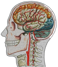
The brain is made up of billions and billions of neurons that have the power to generate specific signals. In return, they can also receive and transmit these signals from their surrounding cells. The word ‘neuron’ is translated into ‘nerve’ and ‘logia’ implies ‘the study of which together forms the term ‘neurology’.
Park Neuroscience Services is equipped with the state of the art diagnostic and treatment infrastructure. All units are supported with the latest radio-diagnosis equipment along with a team of neurologists of national and international repute. Some of the technological interventions being used by the Park Neuroscience Services are:
- Electroencephalogram: An electroencephalogram (EEG) is a test that detects electrical activity in your brain using small, flat metal discs (electrodes) attached to your scalp.
- Electromyography: It is an electro diagnostic medicine technique for evaluating and recording the electrical activity produced by skeletal muscles.
- Sleep Study Lab: A sleep study is an overnight evaluation that records brain activity, eye movements, heart rate, blood pressure, oxygen levels, body movement, and more.
- Ultrasound of the brain : Ultrasound imaging of the head uses sound waves to produce pictures of the brain and cerebrospinal fluid. It is most commonly performed on infants, whose skulls have not completely formed. A transcranial Doppler ultrasound evaluates blood flow in the brain’s major arteries.
- Functional MRI: It is one of the most preferred radiological diagnostic tools that can help diagnose the location, size and position of the brain tumor. The same can also help in detecting the type of the brain tumour and provide clear guidance on the treatment protocol for the same.